


by: Samar Bhat
Today, one of the most daunting challenges physicians face in treating cancer is containing its growth. If a cancerous tumor can be confined to a known region in the body, it would be much easier to treat with conventional methods. The problem is that most, if not all cancerous tissue, upon reaching a certain point in its growth and development, gains a propensity to metastasize, or spread by passage of our very own circulatory systems. But what if there was a way to follow and detect cancer cells, even as they attempt to escape our grasp?
Circulating tumor cells (CTCs) break off a primary tumor, and gain access into the bloodstream by penetrating the thin wall of endothelial cells and supportive mesenchymal tissue. Once in the bloodstream, the tumor cells get lost amongst billions of red and white blood cells, after which they are essentially impossible to track with currently available tools. Without being able to track where these cells travel, we have no real idea of where to search for growing metastases, and therefore no viable strategy for early prevention and treatment which may significantly improve a patient’s prognosis.
Fortunately, a recently patented method developed in the lab of Dr. Amy Herr at UC Berkeley, may be able to solve this problem. In order to fully describe a cell’s phenotype in a way that would be useful for developing treatments and diagnostic tools, we must be able to make precise measurements of its genome, transcriptome, and protein content. Currently, the technology for analyzing the genome and transcriptome (DNA and RNA) of single cells is more sophisticated than that which exists for detecting protein expression at single cell resolution. But Dr. Herr and her colleagues have found a way to analyze these cells at a higher resolution than was ever before possible by focusing their model on the analysis of surface protein expression on single CTCs.
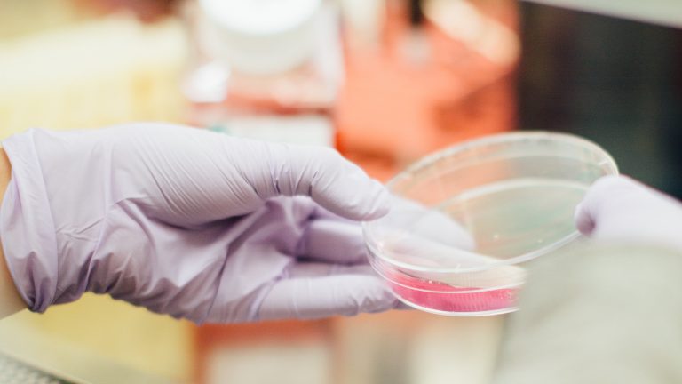
The device’s core technology utilizes microfluidics to perform multiplexed western blotting on patient blood samples. Western blotting is a technique used commonly in molecular biology to isolate and detect proteins of interest within a sample. Thus, by analyzing a set of characteristic proteins known to be found on a tumor cell of interest, western blotting a sample of blood makes it possible to detect these tumor cells in vivo.
In the paper describing this technology, “Profiling protein expression in tumour cells using microfluidic western-blotting,” the authors explain how a protein panel of eight key proteins found on estrogen receptor-positive (ER+) breast cancer cells was used to detect these cells in blood. In the experimental setup, a sample of whole blood is first enriched for large nucleated cells using a “size- and deformability -selective microfluidic tool” to select cells with diameters larger than white blood cells, thereby excluding red blood cells, which are even smaller than WBCs.
These cells are then loaded onto a microfluidic chip coated with a thin layer of polyacrylamide gel patterned with microwells whose diameters are just large enough to accomodate a single cell. When a sample of cells is flowed onto the chip, each cell settles into its own microwell and chemical reagents are added to rupture the cells to release their contents. To probe the sample of cells for the proteins of interest, specific antibodies are added to the wells and are labelled with radioactive molecules. This way, when cancer cells containing specific proteins are bound by the radiolabeled antibodies, they fluoresce and are easily detected by scientists.
Although this experiment was shown to work only on a very specific cell type from a very specific cancer phenotype, it has huge implications for the development of more powerful and efficient techniques for liquid biopsy. When asked about the single most powerful implication of this device in the treatment and diagnosis of cancer, Dr. Herr shared that the device will “hopefully get to the point where it can be used in clinics as a point-of-care diagnostic tool”. However, she qualified her statement by shedding some light on the difficulties of obtaining metrics with absolute precision in biological samples: “as soon as you think you have found a pattern, you quickly find out that the bigger picture is much more complex than you’d previously imagined”.
Liquid biopsy techniques are being rapidly developed as a means to replace traditional biopsy methods, which involve surgery, are more time intensive, and usually involve a greater level of patient distress than a simple blood draw. With improvements in DNA-sequencing, blood collection, and advanced optics-based analytics, the day is fast approaching when patients will be able to monitor themselves with a portable tool.